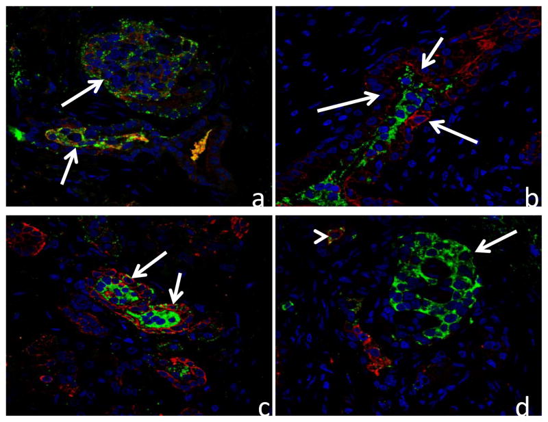
Figure 3.
Confocal microscopy images of pancreas from 3 patients double immunolabeled for insulin (green) and wide-spectrum cytokeratins (red) and nuclear stained with To-Pro 3 (blue). a) Atypical islet-like structures in case #1 showing cells with both insulin and wide-spectrum cytokeratin immunoreactivity (arrows). b) Atypical islet-like structure in case #2 showing insulin positive cells surrounded by a duct-like structure (arrows) in which cells label for wide-spectrum cytokeratins. c) Atypical islet-like structure in case #3 showing insulin positive cells surrounded by a duct-like structure in which cells label for wide spectrum cytokeratins. Arrows demonstrate cells with both insulin and cytokeratin immunoreactivity. d) Normal islet (arrow) in case #3 in which β-cells do not show cytokeratin immunoreactivity. A single atypical cell (arrow head) labels for both insulin and cytokeratins.

