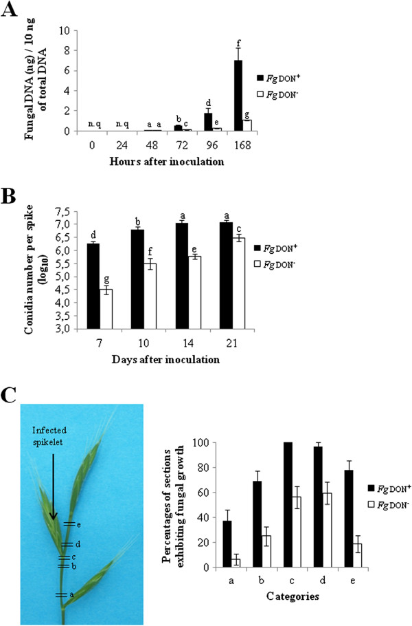
Figure 3.

Estimation of B. distachyon spike (let)s colonization of F. graminearum Fg don + and Fg don - strains . A: Quantification of fungal DNA in infected spikelets (n.q. = not quantifiable, different letters indicate significant differences between conditions; t-test, p-value ≤ 0.01). B: Production of macroconidia on B. distachyon spikes infected by the Fg don + or the Fg don - strain; different letters indicate the significance of differences between conditions (Duncan test, p-value ≤ 0.01). C: Evaluation of B. distachyon rachis colonization by the Fg don + or the Fg don - strain on infected spikes collected 7 dai; left panel: localization of the different rachis sections collected, right panel: quantification of sections presenting out of which fungal growth was observed.
