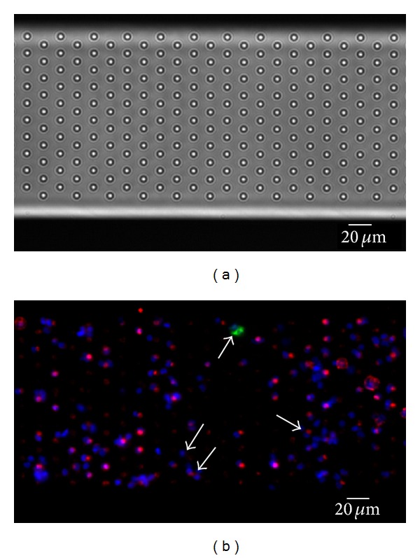
Figure 5.

Cells from CellSearch Waste immunostained on a microsieve. Blood from a lung cancer patient was used for a CellSearch assay. After immunomagnetic selection, part of the sample was discarded by the system and used for filtration on a microsieve with 5 µm pores. Bright-field image of the sieve is shown in (a). (b) shows the sieve with filtered sample. Cells were stained for nucleus (blue), cytokeratin C11 (green), and CD45 (red). Fat arrow points to a CTC, positive in CK. Small arrows point to the absent staining of cells, showing the difficulty of accounting for all cells on the sieve. Image taken on a fluorescence microscope with a 10x (0.45NA) objective.
