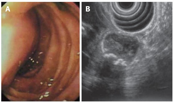
Figure 2.

Endoscopic (A) and EUS (B) findings. By endoscopy the lesion appears as a submucosal mass obstructing the bile ostium and protruding into the duodenal lumen; EUS procedure was impossible to obtain further information because of the cystic lesion large size. The presence of calculi in the lesion was
