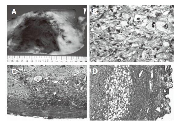
Figure 4.

Resected specimen and histological findings. A: Cutting section of the tumor; B: The main tumor; C: The edge of the tumor(hepatic side); D: The posterior wall of the gallbladder. The cutting section of the tumor appeared whitish solid and included parts of hemorrhage and necrosis and attached to the gallbladder firmly (A) (arrowhead: gallbladder). On histological examination, the tumor was mainly composed of spindle cells with cellular pleomorphism, and included lipoblasts (B). The tumor had a capsule (C), but the capsule annihilated a border of the gallbladder, where these tumor cells were detected in the muscle layer of the gallbladder (D).
