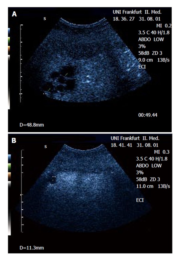
Figure 1.

Demonstration of a focal (multicystic) lesion in a patient with cervix carcinoma using contrast-enhanced ultrasound (CEUS). A: The lesions can be delineated in the portal-venous phase as ‘black spots’ lacking portal-venous enhancement within normally enhanced liver tissue. B: An additional small lesion next to the diaphragm (not visible in native B-mode) was detected by CEUS but not with CT. Biopsy confirmed metastatic disease.
