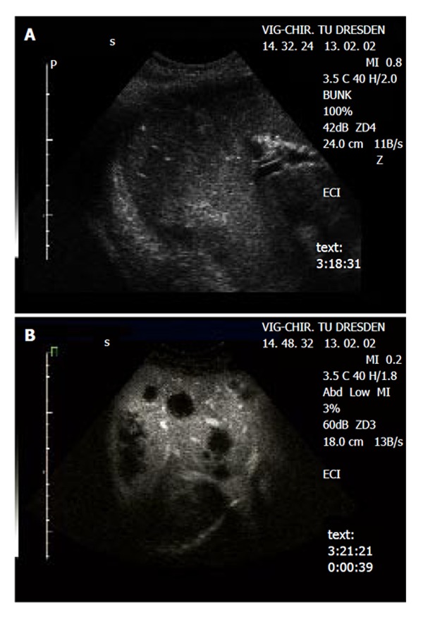
Figure 2.

Detection of metastases in a patient with colorectal carcinoma. A: Native B-mode sonography revealed 3 metastases (segment 6/7) in agreement with CT, MRI revealed 4 metastases (segment 6/7 and 4). B: Contrast-enhanced sonography identified diffuse metastatic disease in both liver lobes. The metastatic lesions are clearly delineated in the portal-venous phase as ‘black spots’, due to the lack of portal-venous blood supply.
