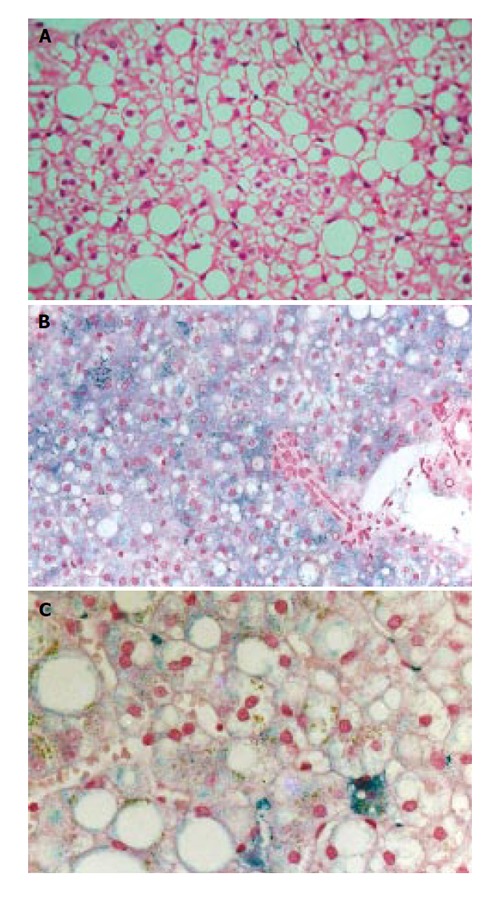
Figure 1.

Case 1 liver histology. A: Hematoxylin and eosin staining shows grade 2 hepatic steatosis; B: Perls’ staining shows grade 1 hepatocyte siderosis with predominant periportal distribution; C: Higher power of Perls’ staining shows clustered Kupffer cell siderosis (right lower field) and also irregular large granular deposits in sinusoidal cells.
