
Figure 4.
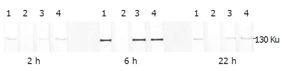
iNOS determination by SDS-polyacrylamide gel electrophoresis and by Western blotting in Kupffer cells stimulated by L. interogans and B. burgdorferi. The 130 ku iNOS protein band was detected by specific polyclonal rabbit anti-iNOS antibodies. The iNOS protein band was evident 2 h after infection. The band peaked 6 h after infection and was still evident 22 h after infection. Lane 1: Kupffer cells stimulated by LPS and interferon-γ (positive control); lane 2: Kupffer cells not stimulated (negative control); lane 3: Kupffer cells stimulated by L.interrogans; lane 4: Kupffer cells stimulated by B.burgdorferi.