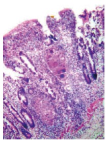
Figure 6.

Histological examination of the biopsies obtained from the area of nodularity and ulceration in the terminal ileum showing noncaseating granulomas consisting of Langhans’ giant cells and epithelioid cells. Note that a cuff of lymphocytes surrounds the granuloma (HE ×80).
