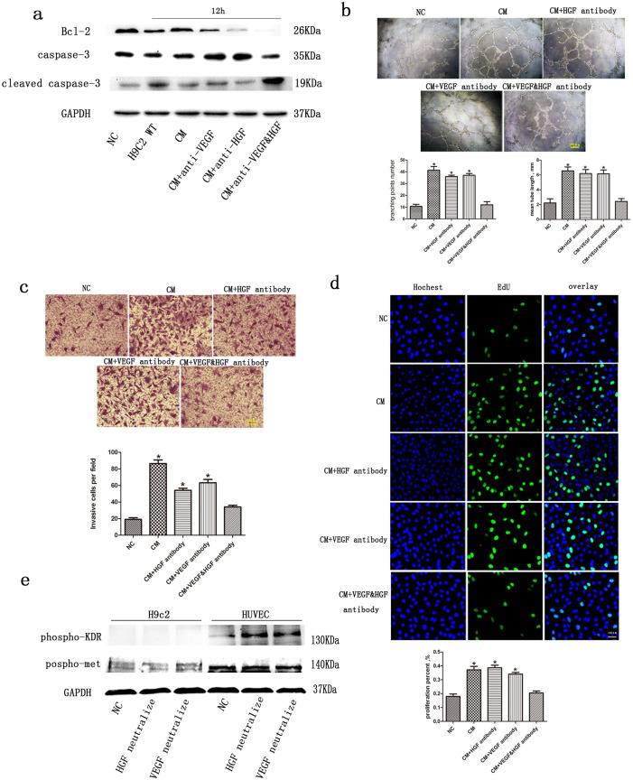
Figure 4. Paracrine and autocrine actions of VEGF and HGF on angiogenesis and cytoprotection.
(a) Representative immunoblots of Bcl-2 and cleaved caspase-3 in H9c2 cells treated with conditioned medium in the presence or absence of neutralizing antibodies after treatment with hypoxia for 12 h. GAPDH served as the loading control. The preparation of conditioned media is described in the Material and Methods section. Concentrated medium (10 μl) from p27kip1-knockdown H9c2 cells treated with hypoxia for 12 h was applied to 1-ml cultures of HUVECs or H9c2 cells. (b) Representative photomicrographs of HUVEC tube formation assays. Quantitative analysis of tube length or branching points after various treatments. *p < 0.05 versus NC (n = 3). (c) Representative photomicrographs of HUVEC migration during transwell assays. Quantitative analysis of migrated cells after various treatments. *p < 0.05 versus NC. (d) Representative photomicrographs of HUVEC proliferation measured using EdU incorporation assays. The numbers of Edu positive cells after various treatments were calculated. *p < 0.05 versus NC. (e) Representative immunoblots of phosphor-KDR and phosphor-Met in both H9c2 cells and HUVECs treated with conditioned medium in the presence or absence of neutralizing antibodies after treatment with hypoxia for 12 h. GAPDH served as a loading control.

