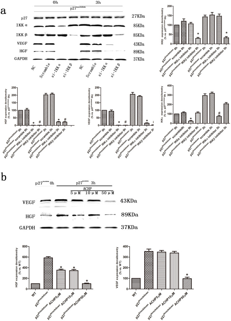
Figure 6. The role of IKK in VEGF and HGF production in cardiomyocytes under hypoxic conditions.
(a) Representative immunoblots of VEGF and HGF in p27kip1-knockdown H9c2 cells treated with IKKα or IKKβ siRNA and then exposed to hypoxia for 3 h. GAPDH served as the loading control. Quantification is shown, *p < 0.05 versus p27 knockdown IKKα/IKKβ inhibitor 0 h; #p < 0.05 versus IKKα/IKKβ inhibitor 3 h (n = 3). (b) Representative immunoblots of VEGF and HGF in p27kip1-knockdown H9c2 cells treated with the pharmacological IKK inhibitor, ACHP, and then exposed to hypoxia for 3 h. GAPDH served as the loading control. Quantification is shown, *p < 0.05 versus p27 knockdown (n = 3).

