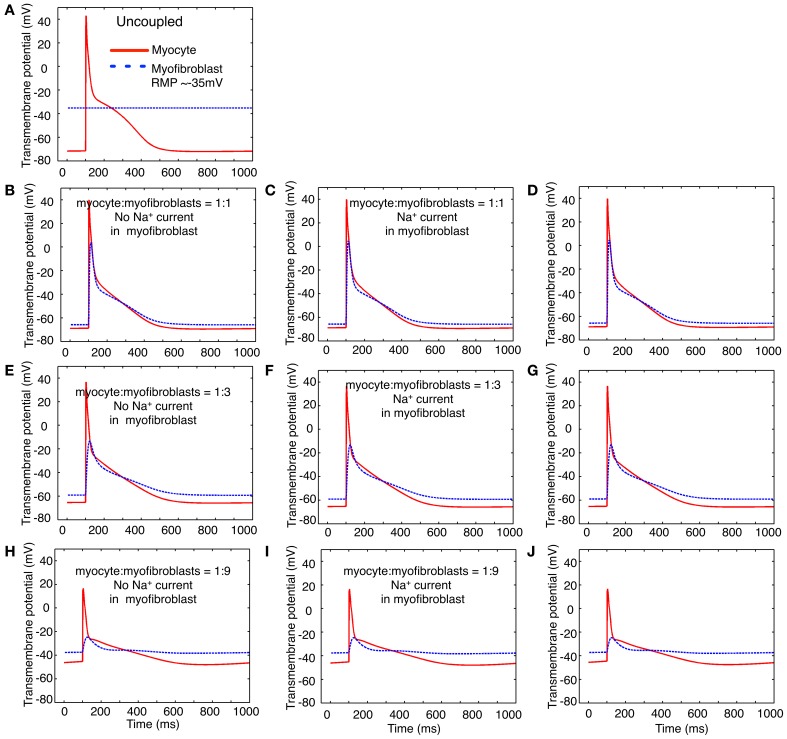
Figure 7.
Illustration of the effects of coupling selected numbers of myofibroblasts to 1 human atrial myocyte using an in silico hybrid mathematical model similar to the one first described by Maleckar et al. (2009a) when assuming a myofibroblast resting potential of −35 or −65 mV. Panel (A) shows the human atrial myocyte action potential (red) and the myofibroblast at a resting potential of −35 mV, prior to these cells being coupled through a linear intercellular conductance of 0.5 nS. The remainder of this Figure is arranged in three rows. Panels (B–D) illustrate responses obtained when the myocyte to myofibroblast ratio is 1:1, panels (E–G) were computed based on a myocyte to myofibroblast ratio of 1:3; and panels (H–J) show responses when myocyte to myofibroblast ratio was 1:9. The arrangement of the vertical columns allow a direct comparison of the electrophysiological effects of (a) there being no Na+ current in any myofibroblast (left column, panels B,E,H); (b) the Na+ current identified in our laboratory and described in the paper being introduced (panels C,F,I); or (c) the Na+ current reported by Chatelier et al. (2012) being added to each myofibroblast (D,G,J). Note that the myocyte action potential causes a significant electrotonic waveform in each myofibroblast at ratios of 1:1 and 1:3. At a coupling ratio of 1:9, the myofibroblasts exert a pronounced depolarizing influence of the myocyte resting potential in these assumed conditions (see Discussion).

