
Fig. 6.
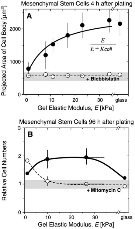
MSC spreading, proliferation, and drug responses. A) Area of MSC cell body as measured on PA gels of different stiffnesses and on glass. Cell area increases with stiffness unless treated with the myosin II inhibitor, blebbistatin. The solid line shows the best fit using the function shown as inset yielding K coll=10 kPa that agrees well with values determined for smooth muscle cells. B) MSC cell numbers relative to the number plated on elastic PA hydrogels. On soft gels, mitomycin C treated cells (open circles) seem to be more proliferative than the untreated control cells (full grey circles), whereas the behavior reverses on very rigid surfaces and on glass, proliferation is suppressed due to strong adhesion to stiffer substrates.