
FIGURE 1. Characterization of the Rdh12 knock-out mice.
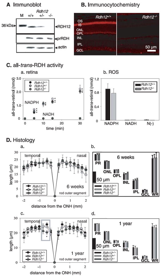
A, immunoblots of Rdh12+/+, Rdh12+/−, and Rdh12−/− retina extracts probed with anti-RDH12, anti-RDH8 (prRDH), and anti-actin polyclonal antibodies. M, molecular mass standards. B, immunocytochemical localization of RDH12 (red) in 8-week-old Rdh12+/+ and Rdh12−/− retina frozen sections. C, all-trans-RDH activity in the retina (NADPH versus NADH) (a) and ROS from Rdh12+/+, Rdh12+/−, and Rdh12−/− mice (b). The bars indicate the S.E. of the mean (n > 5). RDH activities of the retina and ROS (homogenates with the reaction buffer) of mice were assayed by monitoring the production of all-trans-retinol (reduction of all-trans-retinal). The reaction mixture (100 μl of 10 mm sodium phosphate buffer, pH 7.0, containing 100 mm NaCl and 1 mm n-dodecyl-β-maltoside) contained one retina or 5 μg mouse ROS membrane in the presence of NAD(P)H (1 mm) or absence of dinucleotide (N(–)). The reaction was initiated by the addition of 20 μm all-trans-retinal, incubated at 37 °C, and then terminated with 400 μl of CH3OH, and the retinoids were extracted with twice 500 μl of hexane. The hexane solution was analyzed by HPLC using 10% ethyl acetate in hexane to measure the production of all-trans-retinol. D, retina histology of Rdh12+/+, Rdh12+/−, and Rdh12−/− mice. ROS length (in μm) is plotted as a function of distance (in mm) from the optic nerve head (ONH). The ages of mice were 6 weeks (a) or 1 year (c). Quantification of the thickness of different layers of the retina from Rdh12−/−, Rdh12+/−, and Rdh12+/+ mice were measured at 1.25mmsuperior to the optic nerve head. The ages of mice were 6 weeks (b) or 1 year (d). Error bars indicate the S.E. of the mean (n > 3). OS, outer segment; IS, inner segment; ONL, outer nuclear layer; OPL, outer plexiform layer; INL, inner nuclear layer; IPL, inner plexiform layer; GCL, ganglion cell layer; WR, whole retina (*, p < 0.001).