
FIGURE 3. Kinetics of all-trans-retinal reduction and 11-cis-retinal regeneration in Rdh12+/+ and Rdh12−/− mice with RPE65 (Met-450).
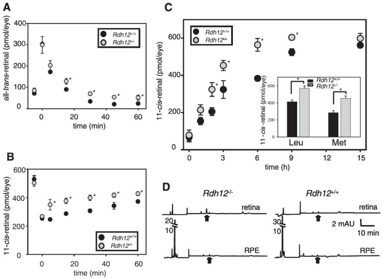
Retinoids were quantified by HPLC on samples collected at different time points after a flash that bleached ~35% of the visual pigment for the pigmented mice. A, changes in the all-trans-retinal levels. B, changes in the 11-cis-retinal levels. Error bars indicate the S.E. of the mean (n > 3). Mice were reared under 12 h/12h dark/light cycle conditions. (*, p < 0.001). C, dark-adapted mice were exposed to background light of 500 cd·m−2 for 24 min (~90% rhodopsin bleach) and returned to the dark until retinoid analysis by HPLC. Amounts of 11-cis-retinal at several time points after the bleach were plotted. Inset, amount of 11-cis-retinal was examined at 3 h of dark adaptation after the light of 500 cd·m−2 for 24 min in Rdh12−/− and Rdh12+/+ mice with Leu and Met at position 450 of RPE65. Error bars indicate the S.E. of the mean (n > 3) (*, p < 0.001). D, dark-adapted mice were exposed to background light of 500 cd·m−2 for 48 min (~98% rhodopsin bleach) and returned to the dark. Retinoid analysis of the dissected retina and RPE was performed separately immediately after the bleach. There was unavoidable cross-contamination of the RPE with retina and vice versa as measured by the presence of retinyl esters in the retinal fraction and 11-cis-retinal in the RPE. Representative chromatograms are shown (n > 3). Black arrows indicate elution times for syn- all-trans-retinyl isomer.