
FIGURE 5. Light induced retinal degeneration under various light exposure durations.
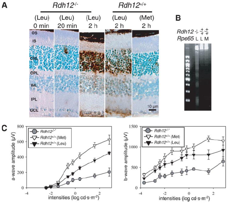
LD was induced in Rdh12−/− and Rdh1+/+ mice with Leu or Met at position 450 of RPE65 mice with dilated pupils by exposure to 10,000 lux of diffuse white fluorescent light as described under “Materials and Methods”. A, apoptotic cells were detected by terminal dUTP nick-end labeling stain after various exposure times. Representative retinal histology 500 μm from the optic nerve head is presented. B, formation of a DNA ladder in agarose gel electrophoresis was examined 24 h after 2 h illumination. L, Leu; M, Met. C, the level of retinal damage was evaluated with full-field ERG 24 h after intense light exposure. Both a- and b-wave amplitudes were attenuated significantly in Rdh12−/− mice expressing RPE65 (Leu-450) compared with Rdh12+/+ mice expressing RPE65 (Met-450) (*, p < 0.001).