
Fig. 1.
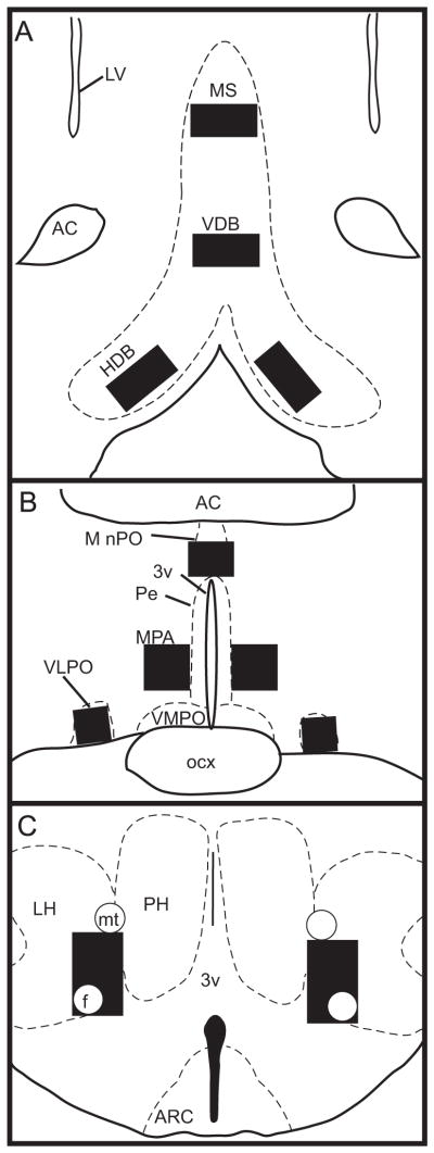
Line drawings depicting location of the MS, VDB, and HDB (A), MnPO, MPA, and VLPO (B), and LH (C). Sampling boxes (black squares) were used for cell counts in all regions as described in Section 4. Anatomical boundaries are based on Paxinos and Watson (1997). 3v: third ventricle; AC: anterior commissure; ARC: arcuate nucleus; f: fornix; LV: lateral ventricle; mt: mammillothalamic tract; Pe: periventricular hypothalamus; PH: posterior hypothalamus; ocx: optic chiasm; VMPO: ventromedial preoptic area.