
Fig. 1.
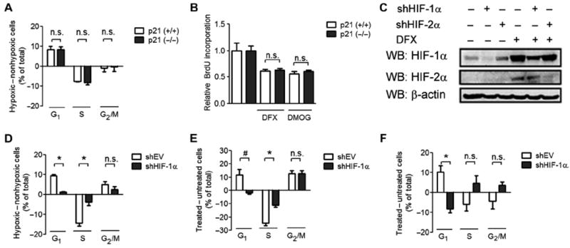
HIF-1α promotes cell cycle arrest that is independent of p21. (A) HCT116 p21 (+/+) and p21 (−/−) cells were exposed to nonhypoxic (20% O2) or hypoxic (1% O2) culture conditions. The difference in the percentage of cells at the indicated stage of the cell cycle between the hypoxic and the nonhypoxic states is shown as means ± SEM; n.s., difference is not significant. n = 3 to 4 independent replicates. (B) HCT116 p21 (+/+) cells (open bar) or p21 (−/−) cells (closed bar) were left untreated or treated with DFX or DMOG and then labeled with BrdU. BrdU labeling was measured, and data were normalized to the untreated control. n = 3 to 4 independent replicates. (C) HCT116 cells were infected with shEV or vector encoding shHIF-1α or shHIF-2α. Cells were treated with DFX or left untreated. Cell lysates were subjected to Western blot (WB) assays using antibodies against HIF-1α, HIF-2α, and β-actin. Data are representative of two independent experiments. (D to F) HCT116-shEV or HCT116–shHIF-1α cells were exposed to 20 or 1% O2 (D), treated with DMOG (E), or treated with DFX (F). The difference in the percentage of cells at the indicated stage of the cell cycle between the treated and the untreated states is shown as mean ± SEM. *P < 0.01; #P < 0.05; n.s., not significant. n = 3 to 4 independent replicates.