
Figure 1.
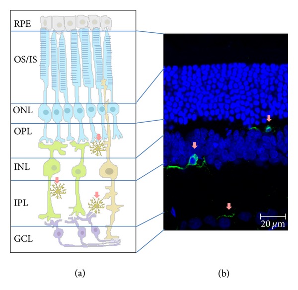
Microglial localization in the retina. Microglial cells in a “surveying” state (pink arrows) in nonpathological conditions are mainly located in the plexiform layers. Retinal layers: OS/IS, outer and inner segments of rods and cones; ONL, outer nuclear layer; OPL, outer plexiform layer; INL, inner nuclear layer; IPL, inner plexiform layer; GCL, ganglion cell layer. Schematic draw of the retinal layers (a) and confocal image from a retinal section where the different layers are depicted (b): nuclear layers (in blue) and microglia cells (in green).