
Figure 3.
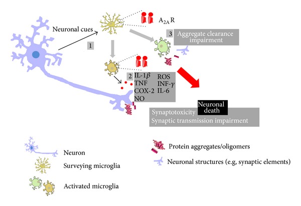
Microglia and neuroinflammation in the brain/retina. Schematic representation of the main inflammatory responses mediated by microglial cells (in yellow) in neurodegenerative conditions. Environment surveillance allows the detection of “pathological” events affecting neurons (in blue-purple); note that appropriate detection of danger signals may also be compromised under these conditions; one of the microglial changes consists in the upregulation of the expression/density of A2AR, as described in several degenerative disorders (1), usually paralleled by morphologic changes and by the release of inflammatory mediators (red circles), both anti- and proinflammatory molecules, that may impact on synaptic transmission, ultimately leading to synaptotoxicity (2); the ability of microglia to phagocytose subcellular components of damaged neurons or protein aggregates, typically present in some degenerative diseases, may also be impaired, further amplifying the cascade of events that lead to cell death/degeneration (3).