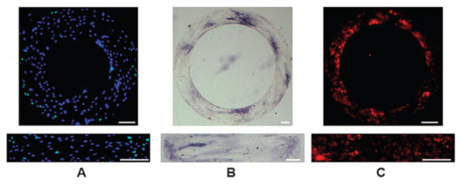
Fig. 2.

Localization of stem cell proliferation and differentiation. (A) Combined fluorescence images of stem cell proliferation on ring and rectangular patterns in growth medium for 1 day (Green: Bromodeoxyuridine (BrdU); Blue: nuclei); (B) Bright field images of Alkaline phosphatase staining (with Fast Blue dye) of human adipose derived stem cells (hASCs) after 3 days incubation in osteogenic medium; (C) fluorescence images of Nile red staining of lipid droplets inside hASCs after cultured in adipogenic medium for 4 days. Scale bars: 100 μm.
