
Fig. 5.
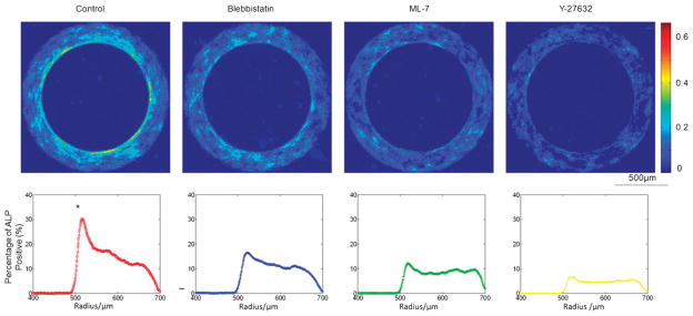
The role of cytoskeleton. Disruption of cytoskeletal tension diminished the patterns of geometrically controlled osteogenic differentiation of hASCs. The frequency maps (top) of ALP activities are shown for stem cells on ring patterns (inner radius: 500 μm and width: 200 μm) after 3 days incubation (from left to right) in osteogenic medium (control) and in osteogenic medium with addition of 10 μM Blebbistatin, 10 μM ML-7, or 2 μ MY-27632. The corresponding cell ALP activities across the radius are shown below the frequency maps. (* significantly different from other groups in the peak ALP activity; p < 0.05).