
Figure 1.
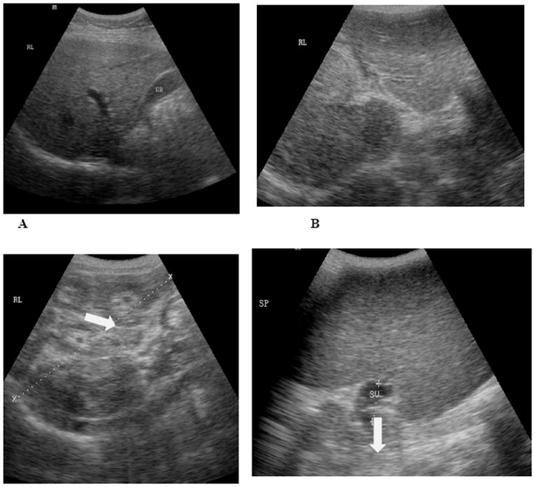
A, B, C, and D are representative ultrasound pictures that are seen in S. japonicum endemic areas. 1A shows a normal liver. 1B shows a liver with Grade II fibrosis (white arrow). 1C is Grade III fibrosis. The “Bull’s Eye” appearance is indicated by the white arrow in this image. Image 1D shows a very large spleen with dilated splenic vein (marked by white arrow).