Figure 1.

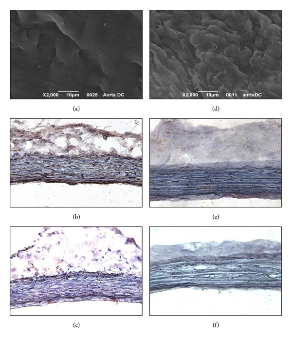
Iliac arteries before (a–c) and after (d–f) the decellularization treatment. (a), (d) SEM micrographs of the luminal sides. Immunoreactivity against MHC I (b, e) and II (c, f) antigens stains brown (magnification ×200).

Iliac arteries before (a–c) and after (d–f) the decellularization treatment. (a), (d) SEM micrographs of the luminal sides. Immunoreactivity against MHC I (b, e) and II (c, f) antigens stains brown (magnification ×200).