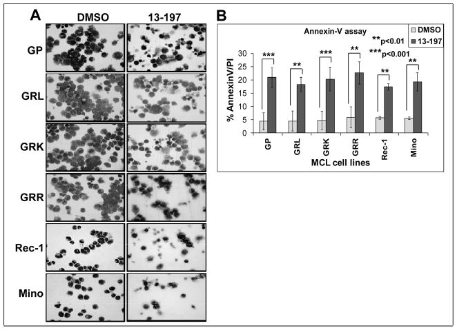
Figure 2. Effect of 13-197 on therapy-resistant MCL cells morphology/apoptosis.
Exponentially growing therapy-resistant and other MCL cells were treated with 10 μM 13-197 for 48 hours. Following treatment, cells were stained with Wright-Giemsa staining using a cytospin preparation. The cells were examined under bright field microscope for the apoptotic cells and images were captured at 40X magnification; A: cytomorphology of different MCL cells indicated above following treatment with 13-197 specifically examining apoptotic bodies; B: Annexin-V apoptosis detection assay was used to access percent of cells undergoing apoptosis in those MCL cell lines following treatment with 10 μM 13-197 for 48 hours. The values represent the means ± SD of three separate experiments.

