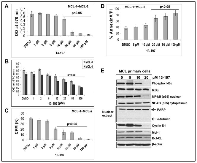
Figure 5. Effect of 13-197 in primary MCL cells.
Primary MCL cells from MCL patients samples (N=4) were cultured in RF-10 medium containing vehicle (DMSO) or indicated concentrations of 13-197 for 24 and 48 hours. Following incubation, cells were subjected for growth/proliferation, apoptosis and western blot analyses. A: MTT assay was used to determine the cell viability in control and 13-197 treated combined cells of two MCL patients (MCL-1 and MCL-2) for 48 hours. The values represent the means ± SD from four wells of the 96-well plates. B: MTT assay was used to determine the cell viability in control and 13-197 treated cells of two additional MCL patients (MCL-3 and MCL-4) for 48 hours. The values represent the means ± SD from four wells of the 96-well plates. C: The proliferation levels in control and treated cells of MCL patients (N=2) were determined using 3[H]-thymidine uptake method following treatment with 13-197 for 48 hours. The values represent the means ± SD from triplicate wells of the 96-well plates. D: Annexin-V apoptosis detection assay was used to access percent of cells undergoing apoptosis in those MCL primary cells (N=2) following treatment with 13-197 for 48 hours. The values represent the means ± SD from triplicate wells of the 6-well plates. E: Following 24 hours of 13-197 treatments, cells were harvested and whole cell lysate or cytoplasmic/nuclear fraction was prepared and subjected to western blot for the expression of indicated molecules of NF-κB pathway. β-actin was used as an internal control in this experiment. This Fig. shows a representative data of two MCL patients.

