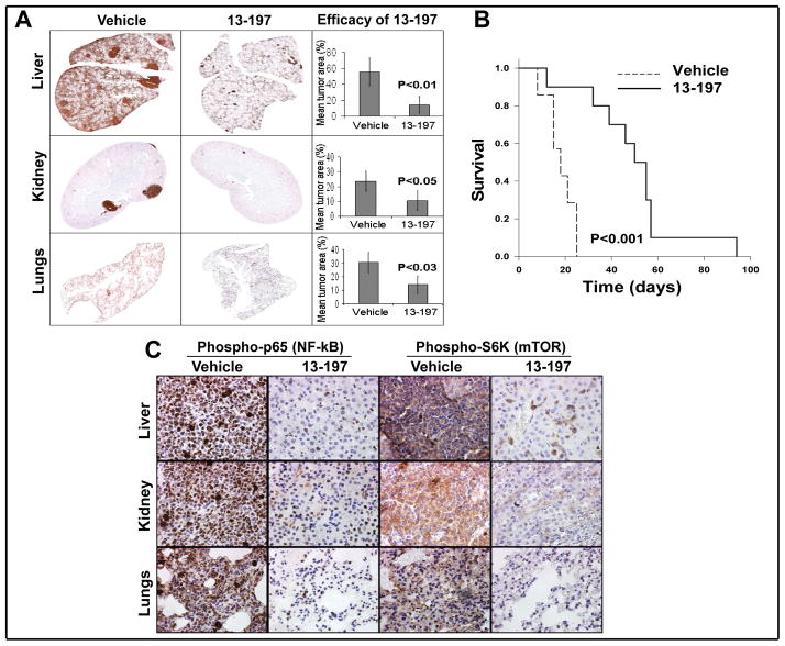
Figure 6. Anti-lymphoma efficacy of 13-197 in a NOD-SCID mouse model in vivo.
NOD-SCID mice bearing therapy-resistant GRL MCL cells were treated with vehicle (cremophor) or 13-197 (120 mg/kg/) for 30 days. A: Immunohistochemical analysis of the liver, kidney, and lungs of MCL bearing NOD-SCID mice following treatment with 13-197. The histological sections from liver, lungs and kidney were stained with anti-CD20 antibody, imaged and quantitated, and analyzed under a digital scanner microscope using Neuroinformatica software. The images of tissue sections from different treatment groups were captured using light microscopy at 10X magnification. B: Kaplan-Meier analyses for the survival of mice using log-rank test. Total number of mice, N=17. C: Immunhistochemical (IHC) analysis for the level of NF-κB (phospho-p65) and mTOR (phospho-S6K) in the liver, kidney, and lungs of MCL bearing NOD-SCID mice following treatment with 13-197. The IHC images were captured using a light microscope at 40X magnification.

