
Figure 1.
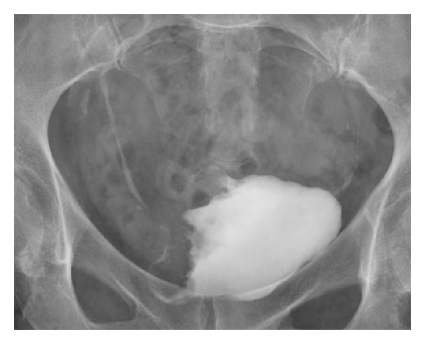
Intravenous urogram (bladder image obtained 15 minutes following contrast administration). There is an infiltrative mass lesion involving the bladder wall on the right. This was confirmed to be a urothelial cell carcinoma following biopsy at cystoscopy.