
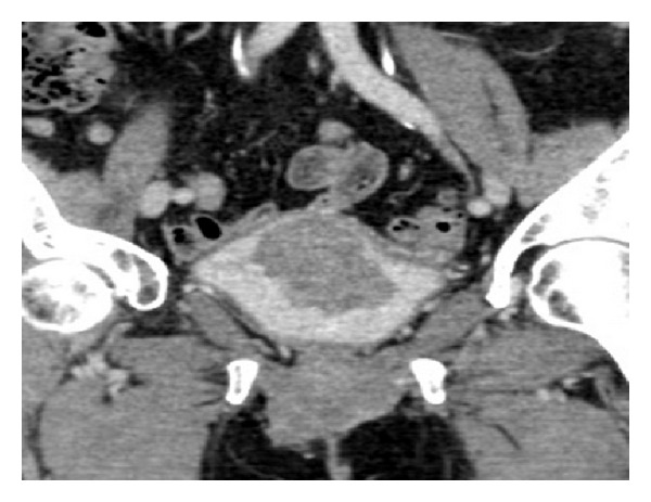
Figure 5.
CT urogram (coronal urographic phase image) demonstrates a large polypoid mass arising from the bladder wall. This was confirmed to be a urothelial cell carcinoma following biopsy at cystoscopy.
Official websites use .gov
A
.gov website belongs to an official
government organization in the United States.
Secure .gov websites use HTTPS
A lock (
) or https:// means you've safely
connected to the .gov website. Share sensitive
information only on official, secure websites.
CT urogram (coronal urographic phase image) demonstrates a large polypoid mass arising from the bladder wall. This was confirmed to be a urothelial cell carcinoma following biopsy at cystoscopy.