
Figure 3.
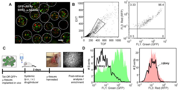
Responsiveness of dual reporter 3D μ-tissues to systemic stimuli in vivo. A,B) 3D GFP+/RFP+ BMEL μ-tissue population characterized by epifluorescence microscopy imaging (A), and quantitative flow analysis along multiple parameters, including time-of-flight (TOF), extinction (EXT), green and red fluorescence (B). C) Timeline of study probing 3D tet-inducible (TetOff-GFP+/RFP+) μ-tissue responsiveness in vivo. 3D μ-tissues are implanted via semi-permeable membrane into the peritoneal cavity of replicate mice, and mice are systemically administered doxycycline (doxy, 2 mg/mL) in 5% sucrose or sucrose only for 2d, prior to membrane harvest and μ-tissue retrieval. Phase micrograph depicts harvested μ-tissues in culture. D) Flow cytometry histograms for green and red fluorescence of n=2,242 3D TetOff-GFP+/RFP+ μ-tissues retrieved from duplicate doxy-treated or control mice. (Scale bars, 200 μm.)