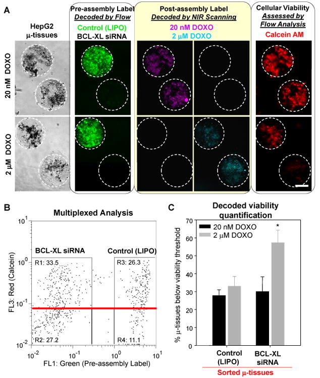
Figure 5.

Multiplex assessment of drug/gene interactions on pooled 3D hepatoma μ-tissues. A) Representative phase and epifluorescence images of lipofectamine (LIPO)-treated or BCL-XL siRNA-treated hepatoma μ-tissues containing green fluorescent or unlabeled pre-assembly tags (left panel). Labeled μ-tissue mixtures were exposed to solutions of doxorubicin (DOXO) containing post-assembly NIR labels (middle panel) and stained with the red calcein AM live dye (right panel). B) Flow cytometry analysis of n=738 pooled hepatoma μ-tissues for green vs. red fluorescence shows real-time decoding of pre-assembly condition (BCL-XL siRNA vs. LIPO) and quantitative viability assessment based on calcein intensity. Enrichment for μ-tissue ‘responders’ was performed by collecting samples with viability below a pre-determined threshold (red line). C) NIR analysis of μ-tissue ‘responders’ enabled identification of post-assembly labels and quantification of the combined effect of BCL-XL siRNA and doxorobucin on hepatoma μ-tissues (see SI Figs. 4, 5 and Experimental). Error bars represent s.d of the mean (n=3). Statistical significance was determined using Student's paired t-test (p<0.05). (Scale bar, 200 μm.)
