Figure 2.

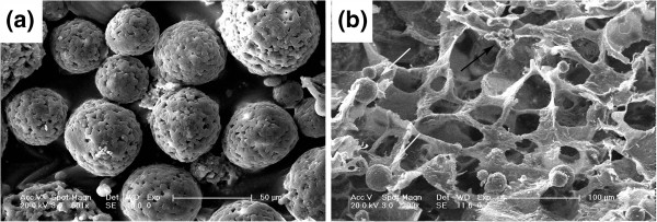
SEM images of chitosan microspheres with ADM (a) and PLGA/nHA scaffold (b) prepared in presence of CMs-ADM. The arrows show the chitosan microspheres (white) and nHA (black) in the scaffold.

SEM images of chitosan microspheres with ADM (a) and PLGA/nHA scaffold (b) prepared in presence of CMs-ADM. The arrows show the chitosan microspheres (white) and nHA (black) in the scaffold.