
Extended Data Figure 4. Nuclear staining of cell cycle matched and mismatched SCNT embryos.
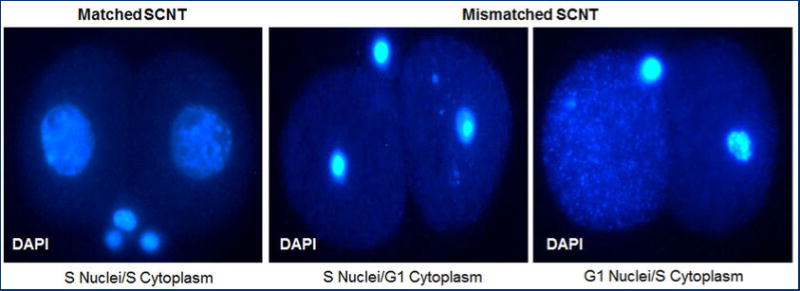
Left panel - expanded nuclei of developmentally competent 2-cell SCNT embryos generated by transfer of S-phase FF nuclei into enucleated S-phase 2-cell embryo. Middle and right panels – Condensed or dispersed nuclei of arrested SCNT embryos generated after cell cycle mismatch between donor nucleus and recipient cytoplasm. Embryos were arrested and apoptotic by the G1/S or S/G2 cell cycle checkpoints, respectively. SCNT embryos were fixed 15 hrs after cleavage.