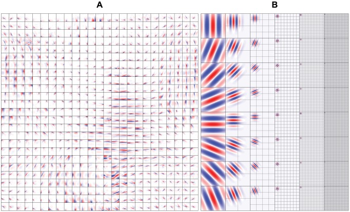
Figure 2. Simple cell receptive fields.
(A) Simple cell receptive fields of the SC model. Each square is of size 32 32 pixels and shows the inverse weights between the input and a simple cell. The receptive fields were topographically organized, spatially localized, oriented and bandpass, similar to those found in the primary visual cortex. (B) Simple cell receptive fields of the GWP model. Each square is of size 128
 128 pixels and shows an even-symmetric Gabor wavelet. The grids show the locations of the remaining Gabor wavelets that were used. The receptive fields spanned eight orientations and six spatial frequencies.

