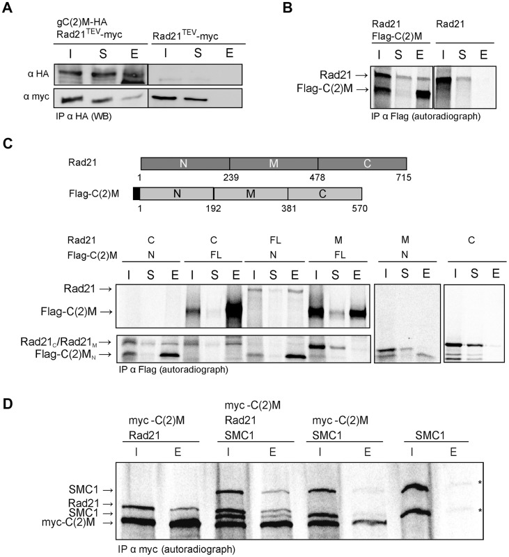
Figure 4. C(2)M physically interacts with Rad21.
(A) Extracts from 0–1.5 h old embryos expressing either h old embryos expressing either gC(2)M-HA together with Rad21TEV-myc or just Rad21TEV-myc were subjected to immunoprecipitation (IP) with mouse anti-HA antibodies. Bound proteins were eluted (E), separated by SDS-PAGE together with input (I) and supernatant after IP (S) samples, and analyzed by western blotting (WB). The blotted samples were probed with anti-HA antibodies to control for immunoprecipitation efficiency and anti-myc antibodies to assess co-precipitation of Rad21TEV-myc. The samples were run on the same gel but not immediately adjacent to each other. Lanes removed from the image are indicated by the vertical black line (B) Full length versions of Rad21 and Flag-epitope tagged C(2)M were synthesized by coupled in vitro transcription/translation (IVT) in the presence of [35S]methionine. IVT reactions were subjected to immunoprecipitation using anti-Flag antibodies. Radioactively labelled proteins were detected by autoradiography. The samples were run on the same gel but not immediately adjacent to each other. Lanes removed from the image are indicated by the vertical black line (C) Schematic of the various Rad21 and Flag-C(2)M fragments assayed for interaction in the coupled IVT-IP experiments. Rad21 fragments were untagged, while all C(2)M fragments were N-terminally fused to 3 copies of the Flag epitope. The proteins were either of full length (FL) or represented the N-terminal part (N), the middle part (M) or the C-terminal part (C) of Rad21 or C(2)M. After IVT-IP using anti-Flag antibodies the samples (I, input; S, supernatant, E, eluate) were separated by SDS-PAGE and radioactively labelled proteins were detected by autoradiography. The migration position of the various fragments is indicated on the left. (D) Coupled IVT-IP of full-length versions of SMC1, Rad21, and myc-C(2)M. After IP using anti-myc antibodies, input (I) and eluate (E) fractions were analyzed. Note that IVT of SMC1 resulted in two protein species, as indicated by asterisks on the right.

