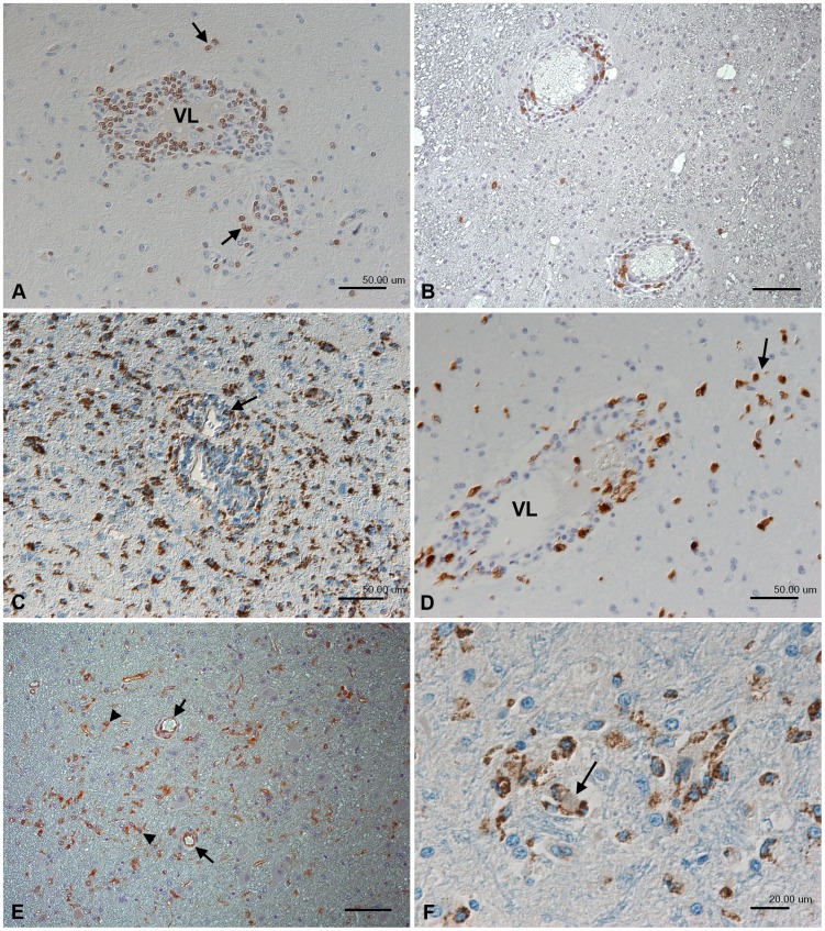
Figure 2. Inflammatory response in the thalamus of rhesus macaques after intranasal inoculation with JEV ((No. 2 (A, B, E) and No. 9 (C, D, F)).
(A) CD3+ T cells dominate the perivascular infiltrates and are present in smaller numbers in the adjacent parenchyma (arrows). VL: vessel lumen. (B) CD20+ B cells represent a minority in the perivascular infiltrates. (C) Staining for CD68 identifies moderate numbers of macrophage/microglial cells within and surrounding the perivascular infiltrates (arrows) and highlights the large number of disseminated activated microglial cells in the adjacent parenchyma. (D) Macrophages in the perivascular infiltrates and the adjacent parenchyma (arrow) also express the myeloid/histiocyte antigen which indicates that they have recently been recruited from the blood. VL: vessel lumen. (E) Activated microglial cells also express major histocompatibility complex (MHC) class II antigen (arrowheads). MHC II is also expressed by vascular endothelial cells (arrows), confirming their activation. (F) Microglial nodule with central degenerate neuron (arrow), surrounded by CD68-positive microglial cells. Indirect peroxidise method, DAB, Papanicolaou's hematoxylin counterstain. Scale bars: A–E = 50 µm; F = 20 µm.

