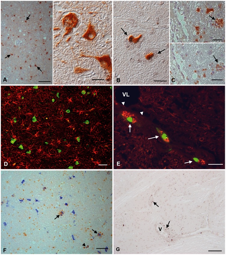
Figure 3. JEV target cells in the thalamus of rhesus macaques after intranasal inoculation with JEV ((No. 7 (A, B), No. 2 (C–G)).
(A) JEV antigen is seen in the majority of neurons (left: arrows). Right: Infected unaltered neurons express viral antigen in both cell body and cell processes. (B) JEV-infected neurons that are surrounded by microglial cells in satellitosis appear shrunken (arrows). (C) Microglial cells in particular in microglial nodules can be JEV-infected (top; arrow) and are identified based on their CD68 expression (bottom; arrow), as demonstrated in a consecutive section. (D) Dual staining for JEV antigen (FITC) and GFAP (Texas red) indicates that JEV does not infect astrocytes. (E) While endothelial cells (arrowheads) were not found to be JEV infected, perivascular macrophages in one animal were found to express JEV antigen (Texas Red); these cells were also undergoing apoptosis, since they were TUNEL-positive (FITC) (arrows). VL: vessel lumen. (F) Dual staining for JEV antigen (Vector Blue) and TUNEL (DAB) shows both the degenerating neurons and surrounding microglial cells in satellitosis undergo apoptosis (arrows). JEV-infected, apoptotic microglial cells (arrowhead) are also observed. (G) Occasional TUNEL-positive, apoptotic lymphocytes (arrows) are present in the perivascular infiltrates. V: vessel. Indirect peroxidase method (A–E, G), Vectastain Elite ABC-Alkaline Phosphatase Kit (F). DAB (A–G), BCIP/NBT blue (F), Papanicolaou's hematoxylin counterstain. Scale bars: A (left) = 100 µm; A (right), C = 25 µm; B, E = 20 µm; D, F, G = 50 µm.

