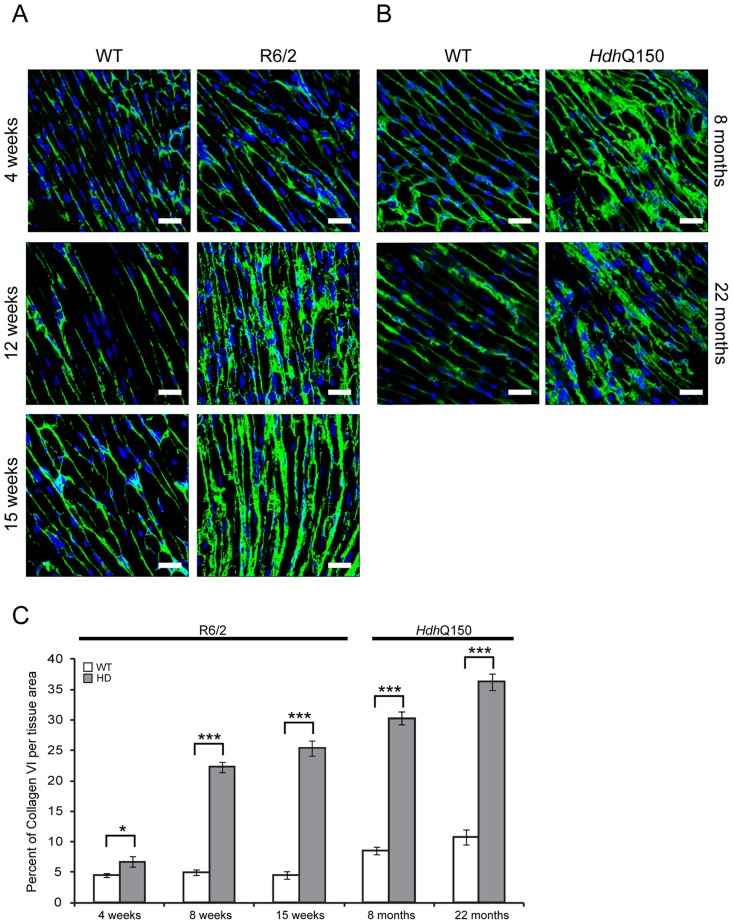
Figure 9. Minor to moderate fibrosis level based on collagen VI deposits in the hearts of HD mouse models.
(A) Representative confocal pictograms of whole heart sections from 4, 12 and 15 week old R6/2 mice and (B) 8 and 22 month old HdhQ150 mice. Fibrosis was detected with the anti-collagen VI antibody (green) and nuclei (blue) were visualised with DAPI. Scale bar 30 μm. (C) Quantification of the collagen VI staining area. Values are mean ± SEM (n = 4). Student's t test: *p<0.05, ***p<0.001.

