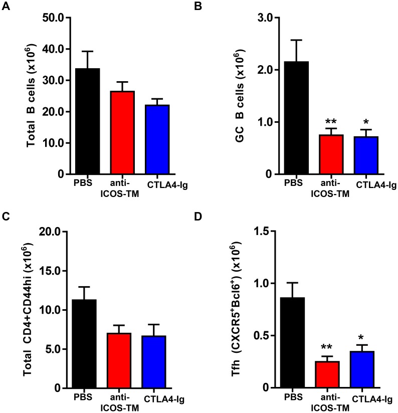
Figure 7. Maintenance of Tfh cells in NZB/W F1 mice requires ICOS signaling.
Female NZB/WF1 mice at 6 months of age were treated with CTLA4-Ig (1 mg mg/mouse) anti-ICOS-TM (1 mg mg/mouse), or PBS at days 0, 2 and 4. Mice were sacrificed and splenocytes were analyzed by FACS at day 7 post first antibody dosing. (A–D) Graphs show number of total B cells (B220+CD19+) in (A), GC B cells (PNA+FAShighIgDlow) in (B), Total CD44high cells in (C) and Tfh cells (CXCR5+PD1high) in (D). **, P<0.01 *, P<0.05. Data are representative of three independent experiments. Bars represent the mean value for each group and error bars are standard error of the mean.

