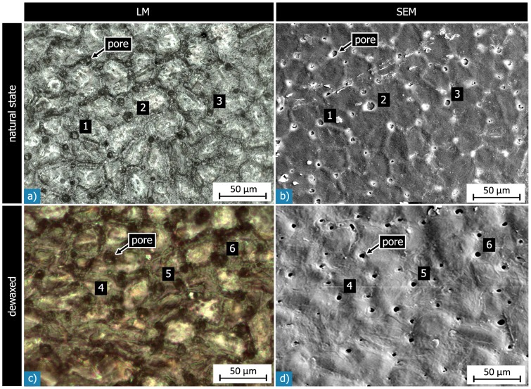
Figure 7. Light (LM) and scanning electron (SEM) micrographs of corresponding sections of the outer surface of the same Macadamia seed coat: a) & b) with natural wax layer, c) & d) after dewaxing.
In both states many pores are seen on the shell’s surface. They appear as dark circular objects in the light micrographs, and are even better visible in the SEM micrographs. The arrows and the numbers denote corresponding pores in the LM and SEM micrographs. The polyhedral surface structure visible in the natural state is less well visible after dewaxing.

