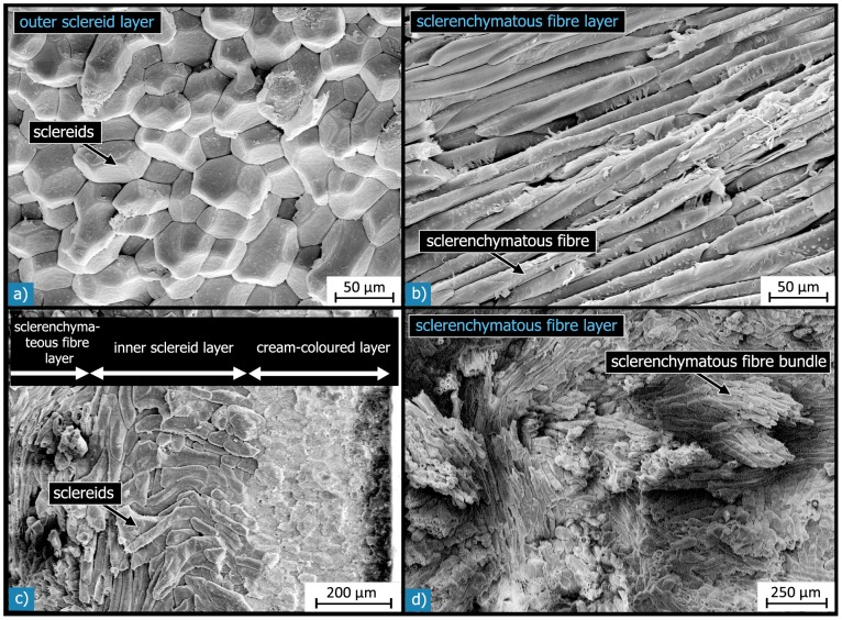
Figure 8. SEM micrographs of the different sclerenchymatous layers.
a) Outer sclereid layer (L2), which is composed of a dense arrangement of polyhedral sclereids; b) the sclerenchymatous fibre layer (L3), which consists of fibrous cells, so-called sclerenchymatous fibres; c) in some regions of the shell, another relatively thin “inner” sclereid layer (L4) was observed, which contains ellipsoidic, kidney- or dumbbell-shaped sclereids; d) the sclerenchymatous fibres are arranged in compact bundles, which are entwined with each other.

