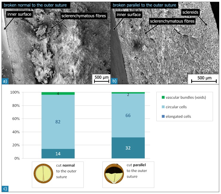
Figure 9. SEM micrographs of Macadamia seed coat fracture surfaces broken normal (a) and parallel (b) to the outer suture.

The “normal” fracture surface (a) is rougher with many sclerenchymatous fibres protruding at different angles. The “parallel” fracture surface (b) is smoother because sclerenchymatous fibres are mainly orientated parallel to the fracture surface. The diagrams in c) show the area fractions of different cell shapes within the sclerenchymatous tissue for sections cut normal or parallel to the outer suture.
