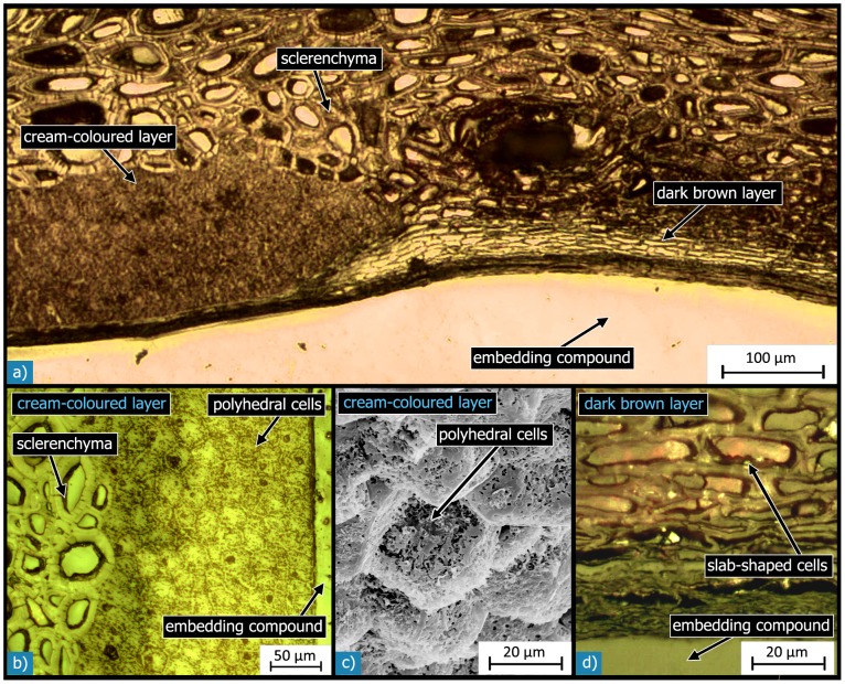
Figure 10. Microstructure of the inner layers and the interfaces between them and to the adjacent layers: Light micrographs of polished sections show the sclerenchymatous and the cream-coloured (L5.1; a, b) and dark brown (L5.2; a, d) inner layers and the interfaces between the different layers.
The cream-coloured layer (a, b) is composed of polyhedral cells with thin cell walls. The SEM micrograph in c) of a fracture surface shows the fine and fibrous microstructure of the cells in the cream-coloured layer. The light micrograph in d) shows the dark brown layer, which is composed of slap-shaped cells with thickened cell walls.

