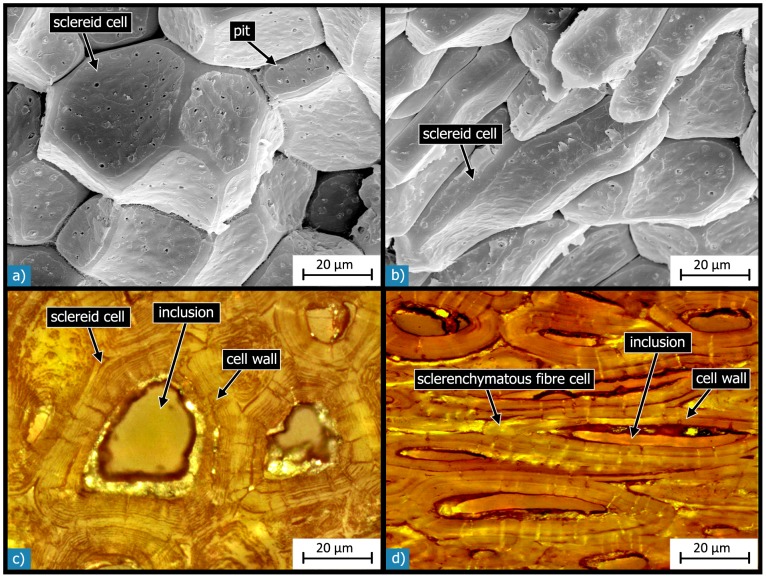
Figure 11. Microstructure of the cells:
SEM micrographs of cells in the outer sclereid layer show that they have an isodiametric shape near the outer surface (a) and a more and more ellipsoidal shape with increasing distance from the shell’s outer surface (b). Light micrographs of polished sections show the structural composition of sclereid cells (c) and of sclerenchymatous fibre cells (d), which have a similar microstructure. Both types of cells have thickened and lignified cell walls and a less dense inclusion within their lumen.

