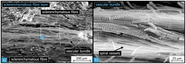
Figure 12. SEM micrographs of vascular bundles within the sclerenchymatous fibre layer:

the vascular bundles are surrounded by sclerenchymatous fibres (a). Each bundle consists of many tube-like cells, so-called spiral vessels and tracheids (b).

the vascular bundles are surrounded by sclerenchymatous fibres (a). Each bundle consists of many tube-like cells, so-called spiral vessels and tracheids (b).