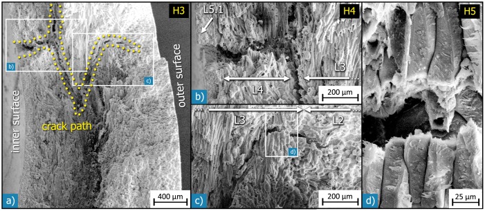
Figure 13. SEM micrographs showing a deflected and branched crack path on different length-scales:
a) fracture surface of an entire Macadamia seed coat after loading in compression; crack deflection took place in three dimensions, as the topography of the fracture surface and the secondary cracks visible on it show. A magnified view of the crack part near the inner surface shows that the crack was deflected at the interface between the inner sclereid layer (L4) and the sclerenchymatous fibre layer (L3). c) The secondary crack stops at the interface between the sclerenchymatous fibre layer (L3) and the outer sclereid layer (L2). d) In the main fracture surface, the crack mostly follows the interfaces between the cells; in this area, however, the secondary crack fractured some sclerenchymatous fibres vertical to their long axis.

