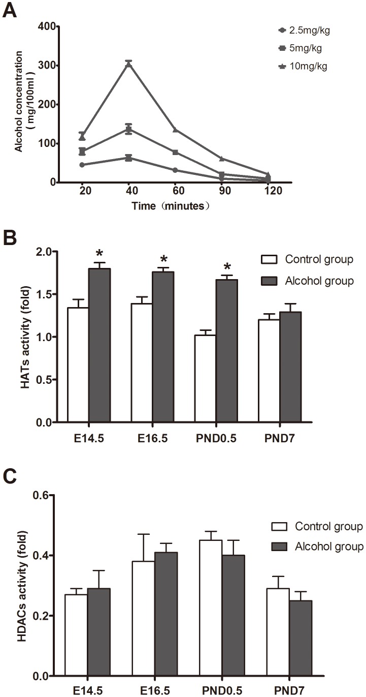
Figure 1. Effects of alcohol exposure on activities of HAT and HDAC.
To analyze the impact of alcohol on activities of HAT and HDAC in cardiac tissues, different doses of alcohol were used to choose optimal exposure dose in pregnant mice. The blood-alcohol concentration (A) after gavaging with different doses of 56% ethanol in mice (n = 6). The alcohol stress (56%) increases HAT activity (B) in E14.5, E16.5 and PND0.5, while it remained unchanged in PND7 and no any effects observed on HDAC activity (C) in myocardial tissues. *: P<0.05 vs. control group (n = 9). E14.5: embryo 14.5 day, E16.5: embryo 16.5 day, PND0.5: postnatal day 0.5, PND7: postnatal day 7.

