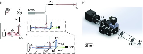
Fig. 1.

Combination optical coherence tomography high resolution fluorescence (OCT/HRF) system. (a) Schematic of HRF imaging integrated within the sample arm of a spectral domain OCT (SD-OCT) system. SLD: superluminescent diode source, C: fiber-optic circulator, PC: polarization controller, L: lens, RM: reference mirror, DG: diffraction grating, LSC: line scan camera, LED: LED excitation source, ex: excitation filter, D: dichroic, em: emission filter, CCD: charge-coupled device camera. (b) Rendering of the OCT/HRF handpiece, drawn to scale. Scale bar is 25 mm.
