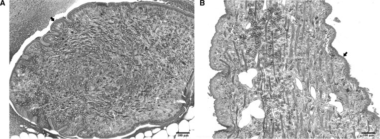
Figure 1.
Representative plerocercoids detected in the paraffin-embedded sections used for molecular identification. (A) A plerocercoid (arrow) isolated from the subcutaneous nodule of the patient 4 (THA-Se4); (B) a plerocercoid (arrow) detected in the spinal cord from the patient 8 (THA-Se8). The sections were stained with hematoxylin-eosin. Scale bar = 100 μm.

